Visual Pathways
- Concentrated at the outer edges of the retina and used in peripheral vision, rods are photoreceptor cells in the retina of the eye that can function in less intense light than the other type of visual photoreceptor, cone cells.
- Cones are one of two types of photoreceptor cells in the retina of the eye. They are responsible for color vision and function best in relatively bright light, as opposed to rod cells, which work better in dim light.
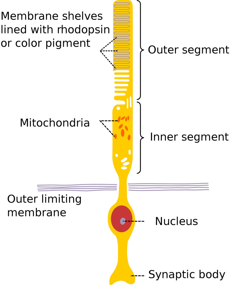
Anatomy of a rod cell.
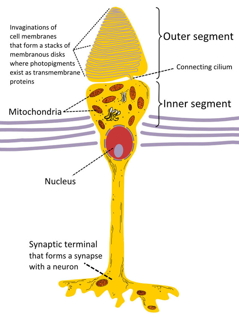
Anatomy of a cone cell.
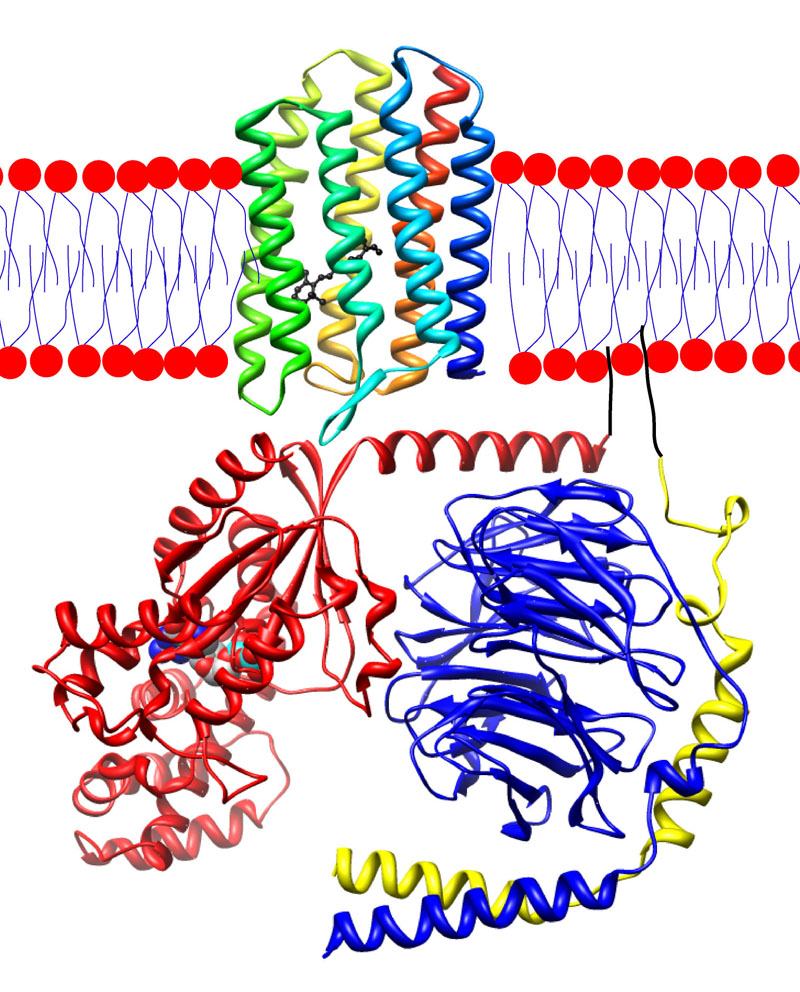
Rhodopsin.
- Rhodopsin, also known as visual purple, is a light-sensitive receptor protein. It is a biological pigment in photoreceptor cells of the retina, the primary pigment found in rod photoreceptors.
- Opsins are a group of light-sensitive proteins found in photoreceptor cells of the retina. Five classical groups are involved in vision, mediating the conversion of a photon of light into an electrochemical signal, the first step in the visual transduction cascade.
- Retinal is a polyene chromophore, bound to proteins called scotopsins and photopsins. It is the chemical basis of animal vision. When it absorbs a photon it undergoes cis-trans isomerization.
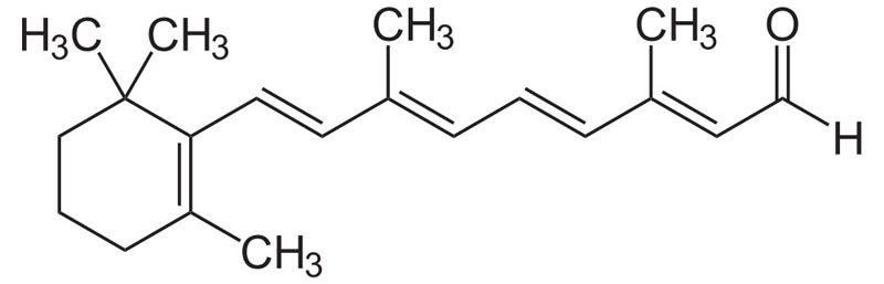
All-trans retinal.
- A type of neuron which has two extensions, bipolar cells are specialized sensory neurons for the transmission of special senses. As such, they are part of the sensory pathways for smell, sight, taste, hearing and vestibular functions.
- Amacrine cells are inhibitory interneurons in the retina. They interact with retinal ganglion cells and/or bipolar cells.
- Horizontal cells are the laterally interconnecting neurons having cell bodies in the inner nuclear layer of the retina of vertebrate eyes. They help integrate and regulate the input from multiple photoreceptor cells.
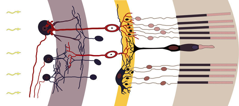
Rods, cones and nerve layers in the retina. The front (anterior) of the eye is on the left. Light (from the left) passes through several transparent nerve layers to reach the rods and cones (far right). A chemical change in the rods and cones send a signal back to the nerves. The signal goes first to the bipolar and horizontal cells, then to the amacrine cells and ganglion cells, then to the optic nerve fibres. The signals are processed in these layers. First, the signals start as raw outputs of points in the rod and cone cells. Then the nerve layers identify simple shapes, such as bright points surrounded by dark points, edges, and movement.
- The optic disc is the point of exit for ganglion cell axons leaving the eye. Because there are no rods or cones overlying this area, it corresponds to a small physiological blind spot in each eye.
- The optic nerve is a paired nerve that transmits visual information from the retina to the brain.
- The optic chiasm is the part of the brain where the optic nerves partially cross. It is located at the bottom of the brain immediately below the hypothalamus.
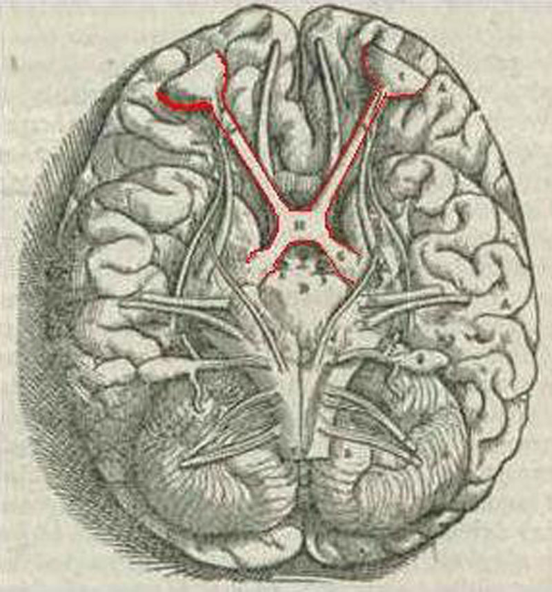
Brain viewed from below showing the visual pathway with optic chiasm.
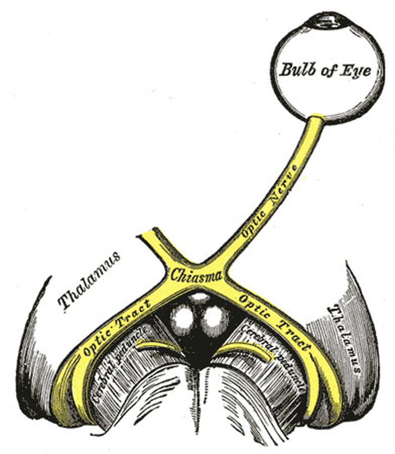
The left optic nerve and the optic tracts.
- The optic tract is a part of the visual system in the brain. It is a continuation of the optic nerve that relays information from the optic chiasm to the ipsilateral lateral geniculate nucleus, pretectal nuclei, and superior colliculus.
- The main central connection for the optic nerve to the occipital lobe, the lateral geniculate nucleus is a relay center in the thalamus for the visual pathway. It receives a major sensory input from the retina.


