Hearing - Structure of the Ear
- The auricle is the visible part of the ear that resides outside of the head. It is also called the pinna.
- The eardrum, or tympanic membrane, is a thin, cone-shaped membrane that separates the external ear from the middle ear.
- The ossicles are three bones in either middle ear that are among the smallest bones in the human body. They serve to transmit sounds from the air to the fluid-filled labyrinth (cochlea).
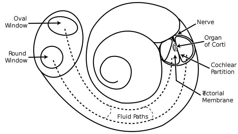
- The oval window is a membrane-covered opening which leads from the middle ear to the vestibule of the inner ear. It serves as the intersection of the middle ear with the inner ear, and is directly contacted by the stapes.
- The round window is one of the two openings from the middle ear into the inner ear. It vibrates with opposite phase to vibrations entering the inner ear through the oval window allowing fluid in the cochlea to move.
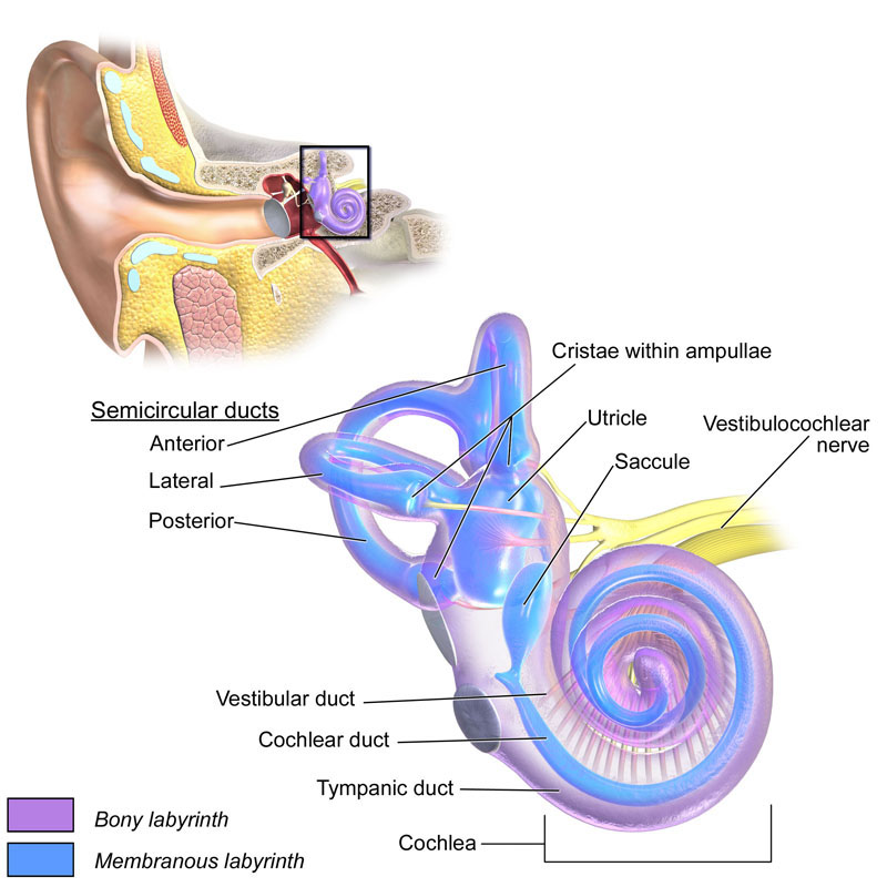
- The cochlea is the auditory portion of the inner ear. It is a spiral-shaped cavity in the bony labyrinth.
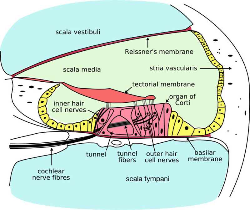
- The organ of Corti, or spiral organ, is the receptor organ for hearing and is located in the mammalian cochlea.
- The basilar membrane within the cochlea of the inner ear is a stiff structural element that separates two liquid-filled tubes that run along the coil of the cochlea.
- Hair cells are the sensory receptors of both the auditory system and the vestibular system in all vertebrates, which through mechanotransduction, detect movement in their environment.
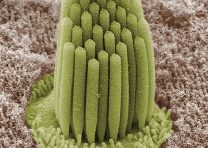
Hair cells.
- In the inner ear, stereocilia are the mechanosensing organelles of hair cells, which respond to fluid motion in numerous types of animals for various functions, including hearing and balance.
- Endolymph is the fluid contained in the membranous labyrinth of the inner ear.
- The tectorial membrane is one of two acellular gels in the cochlea of the inner ear, the other being the basilar membrane.
- Perilymph is an extracellular fluid located within the cochlea in two of its three compartments.


