Human blood smear: a - erythrocytes;...
Electrophoresis of serum proteins
The structure of human serum albumin ...
Human red blood cells
From left to right: erythrocyte, thro...
Blood cell lineage
A scanning electron microscope image ...
Diagram that shows the development of...
Diagram including some of the importa...
Reticulocyte
Erythrocyte
Structure of hemoglobin. The protein ...
Space-filling model of Heme B
Structure of porphine, the simplest p...
Space-filling model of porphyrin
Heme synthesis—note that some reactio...
Structure of Heme B
Heme A Heme A is synthesized from Hem...
The histidine bound haem group of suc...
An example of the globin fold, the ox...
Ribbon diagram of erythrocytic methem...
Animation - Transition between T and ...
Animation - Binding and release of li...
The sigmoidal shape of hemoglobin's o...
Oxyhaemoglobin Dissociation Curve. Do...
2,3-Bisphosphoglycerate
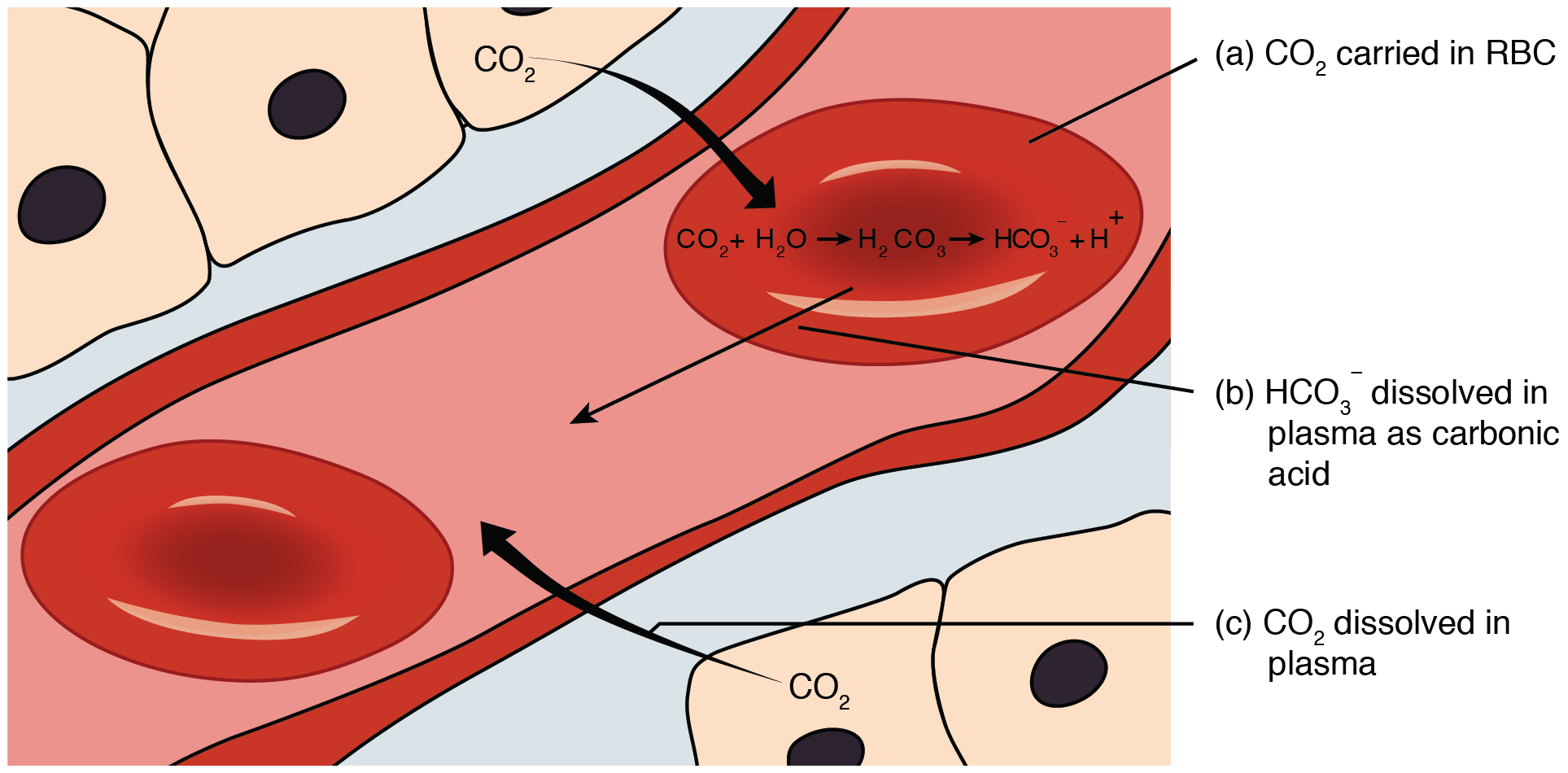
Carbon dioxide transport.
Carbonic anhydrase
Close-up rendering of active site of ...
Ribbon diagram of human carbonic anhy...
Fetal hemoglobin protein structure
The oxygen saturation curve for fetal...
Sickle-shaped red blood cells
A single amino acid change in sickle ...
Distribution of the sickle cell trait
Distribution of Malaria
Sickle-cell disease is inherited in t...
Platelets
Von Willebrand factor
Coagulation factor II (thrombin)
The coagulation cascade.
Blood Coagulation (Thrombin) Pathway,...
Tissue factor
Tissue factor activation
Factor XIII crosslinks fibrin
The Protein C Anticoagulant Pathway: ...
Antithrombin dimer
Fibrinolysis (simplified). Blue arrow...
Crystal structure of the protease dom...
Plasminogen catalytic domain
1PMK Plasminogen Kringle 4
Urokinase
Plasminogen activator inhibitor-1
ABO blood group antigens present on r...
Diagram showing the carbohydrate chai...
A and B are codominant, giving the AB...
Blood group O positive: neither anti-...
Result: Blood group B negative: anti-...
Schematic depicting how the RAAS work...


