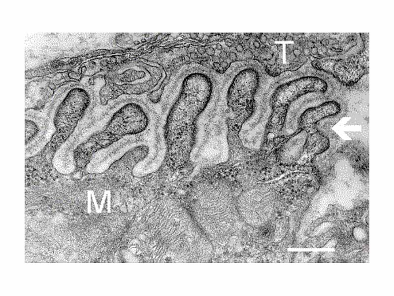
Electron micrograph showing a cross section through the neuromuscular junction. T is the axon terminal, M is the muscle fiber. The arrow shows junctional folds with basal lamina. Postsynaptic densities are visible on the tips between the folds. Scale is 0.3 µm.
Click this LINK to visit the original image and attribution information. Right click on the image to save the 800px teaching JPEG.