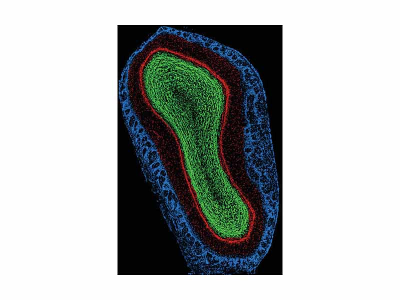
Coronal image of mouse main olfactory bulb cell nuclei. Blue - Glomerular layer; Red - External Plexiform and Mitral cell layer; Green - Internal Plexiform and Granule cell layer. Top of image is dorsal aspect, right of image is lateral aspect. Scale, ventral to dorsal, is approximately 2mm.
Click this LINK to visit the original image and attribution information. Right click on the image to save the 800px teaching JPEG.